Treatment for Glaucoma

Get the Care You Need

Early detection and treatment of glaucoma are essential to prevent permanent visual loss. While some cases of glaucoma can be controlled with medication, more advanced cases may require surgery.
The leading cause of Blindness
About Glaucoma

Glaucoma is a very misunderstood disease. Often, people don't realize the severity or who is affected. Glaucoma can cause irreversible blindness if it is left untreated. Everyone is at risk for glaucoma from babies to senior citizens.

Causes

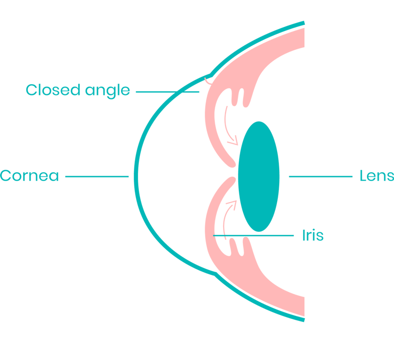
Glaucoma is the leading cause of permanent irreversible blindness. Glaucoma occurs due to fluid building up in the eye as it does not have space to leave the eye. This leads to increased intraocular pressure causing damage to the optic nerve.

Result

The optic nerve is made of more than a million tiny nerve fibers - like an electric cable made up of many small wires. As these nerve fibers die, you will develop loss in peripheral vision. You won't notice blind spots until most of your optic nerve fibers have died. If all of the fibers die, you will become blind.

UNDERSTANDING THE TYPE, PHASE AND TREATMENT FOR GLAUCOMA
Open Angle Glaucoma

Often, open angle glaucoma is hereditary, and the patient is asymptomatic until late in the disease process when poor peripheral vision becomes apparent. This is the most common type of glaucoma. It happens gradually, where the eye does not drain fluid (like a clogged drain). As a result, eye pressure builds and starts to damage the optic nerve. This type of glaucoma is painless and causes no vision changes at first. Some people can have optic nerves that are sensitive to normal eye pressure. This means their risk of getting glaucoma is higher than normal. Regular eye exams are important to find early signs of damage to their optic nerve.

Signs and symptoms of an open angle glaucoma:
  • Reduced vision and loss of peripheral vision
  • Swollen or bulging cornea
  • Advanced stage of pupil dilation to a medium size that doesn't change with increasing or decreasing light
  • Redness in the white of the eye
  • Tunnel vision in the advanced stages
Glaucoma Specialist in Ahmedabad
Angle Closure Glaucoma

The disease is most commonly found among the elderly and those who have small eyes. Known as angle-closure glaucoma or narrow-angle glaucoma, this type occurs when an individual's iris is very close to the drainage angle in their eye. The iris can end up blocking the drainage angle. You can think of it like a piece of paper over a sink drain. When the drainage angle gets completely blocked, eye pressure rises very quickly. This is called an acute attack. It is a true eye emergency, and you should call your ophthalmologist right away or you might go blind.

Many people with angle-closure glaucoma develop it slowly. This is called chronic angle-closure glaucoma. There are no symptoms at first, so they don’t know they have it until the damage is severe or they have an attack. However, 1 out of 3 people (30%) with angle-closure will have a sudden blockage, causing an attack.

Signs and symptoms of an angle closure glaucoma:
 
  • Sudden Blurry Vision
  • Severe Eye Pain
  • Frequent Headaches
  • Upset Stomach & Nausea
  • Vomiting
  • You see rainbow-colored rings or halos around lights
https://www.shivjyotieyehospital.com/wp-content/uploads/2023/12/image2-3.png
Glaucoma Eye disorders leading to progressive damage

If untreated or uncontrolled, glaucoma first causes peripheral vision loss and eventually can lead to blindness. Globally, glaucoma is the second leading cause of blindness (behind cataracts), according to the World Health Organization. Unfortunately, some people have a higher than normal risk of getting glaucoma. If you fall in to any category below you are at a higher risk of developing glaucoma:

If you have a family with history of Glaucoma

If your age is above 40 years

If you feel high pressure on eyes

If you are farsighted or nearsighted

If you are suffering with high blood pressure

If you have uncontrolled diabetes

If you have a history of an eye injury

A detailed examination for
better diagnostics

The only way to diagnose glaucoma is with a complete eye exam. A glaucoma screening that only checks eye pressure is not enough to check for glaucoma. During Glaucoma evaluation, your ophthalmologist will:

  • Measure your eye pressure
  • Inspect your eye’s drainage angle
  • Examine your optic nerve for damage
  • Test your peripheral (side) vision (Perimetry)
  • Take a picture or computer measurement of your optic nerve (Glaucoma - OCT)
State of the Art Technology for
Remarkable Post Operative Results
TOPCON MAESTRO (OCT)

It follows a non-contact high-resolution imaging of the optic disc for the early diagnosis of glaucoma.

MEDMONT PERIMETER

It helps in the Visual Field Testing for diagnosis of glaucoma and comfortable for the patient to get better results.

PREVENT VISION LOSS WITH EFFECTIVE GLAUCOMA TREATMENT
Non-Invasive Treatments

Conservative treatments such as eye drops and oral medications can be used in the early stages of glaucoma to control the amount of fluid in the eye and improve the outward flow. Medications for glaucoma are in the form of eye drops or tablets, as per the requirements. If your eye doctor has advised any eyedrops for glaucoma do not stop or change them without his/her permission. Glaucoma drops are usually advised for lifetime and hence one must not miss his/her daily dose to control the pressure.

Due to long term use of these eye drops, they can cause minor side effects which can be noticed after a long time. They are:

  • A stinging or itching sensation
  • Red eyes or red skin around the eyes
  • Dry mouth
  • Eyelash growth
  • Darkness in the skin around your eyes
Surgical Procedures for
Severe Cases

Surgery can alleviate severe drainage issues by manually creating a new path or placing an implant. Surgical treatment is advised when pressure is not controlled medically. Glaucoma can be safely treated at Shiv Jyoti Eye Hospital using advanced surgical treatment options below:

This is for people who have open-angle glaucoma. The eye surgeon uses a laser to make the drainage angle work better. That way fluid flows out properly and eye pressure is reduced.
This is for people who have angle-closure glaucoma. The ophthalmologist uses a laser to create a tiny hole in the iris. This hole helps fluid flow to the drainage angle.
As shown here, a probe is used to make way for the fluid to drain and thus reduce the pressure.
This is where your eye surgeon creates a tiny flap in the sclera (white of your eye). This will also create a bubble (like a pocket) in the conjunctiva called a filtration bleb. It is usually hidden under the upper eyelid and cannot be seen. Aqueous humor will be able to drain out of the eye through the flap and into the bleb. In the bleb, the fluid is absorbed by tissue around your eye, lowering eye pressure.
Your ophthalmologist may implant a tiny drainage tube in your eye. It sends the fluid to a collection area (called a reservoir). Your eye surgeon creates this reservoir beneath the conjunctiva (the thin membrane that covers the inside of your eyelids and white part of your eye). The fluid is then absorbed into nearby blood vessels. This is advised for advanced stage Glaucoma.
Effective Management of Glaucoma Is Possible with the Right Treatment

Glaucoma is the second leading cause of blindness. If you are experiencing glaucoma symptoms or have neglected to undergo regular eye exams, contact Shiv Jyoti Eye Hospital to schedule an evaluation. Timely treatment can help control your condition and prevent vision loss.

Chat With Us

    Chat With Us