Cornea

the facts you must know

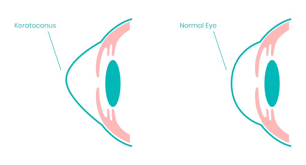
Cornea is the outer covering of the eyeball. Protecting structures within the eye is the primary purpose of this coat. In addition to acting as a structural barrier, the cornea also protects the eye from infection. Combined with the tear film, it provides a proper anterior refractive surface for the eye. The cornea contributes two-thirds of the eye's refractive power.

Corneal Ulcer

A corneal ulcer is an open sore on the cornea, the thin clear layer that covers your iris (the colored part of your eye). It's also called keratitis.
Symptoms of Corneal Ulcer

Corneal ulcers are a type of inflammation of the cornea. The most common causes of corneal ulcers are eye injury, dirty contact lenses, exposure to viruses or dirty water. They are typically caused by infection, but can also be caused by injury.

Common symptoms of corneal ulcer are:

  • Redness
  • Severe pain
  • The feeling that something is in your eye
  • Tears
  • Pus or thick discharge from your eye
  • Blurry vision
  • Pain when looking at bright lights
  • Swollen eyelids
  • A round white spot on your cornea
Treatment of Corneal Ulcer

The treatment for corneal ulcers depends on the cause of the infection. The use of antibacterial and antifungal eye drops can treat diseases caused by bacteria or fungi. You can use eye drops that contain antiviral properties for ulcers caused by viruses. Although these treatments do not cure the virus, it may return in the future. Corneal ulcer can be caused due to bacteria, fungi or viruses. Once diagnosed, a specimen of the ulcer is taken and examined by the microbiologist to know the organism involved. Thus, once the organism is known, treating accordingly gives fast recovery in early stages. If not treated early, infection may spread inside the eye as well further leading to visual loss. Hence, such diseases must be treated as early as possible. Viral keratitis, once treated, never heals completely. If not taken care of, it may occur again.

The picture shows the corneal ulcer with a positive fluorescein stain. Mostly done to confirm the diagnosis of corneal ulcer. (greenish yellow part stained due to loss of first layer of cornea in that area)

With immediate treatment, most ulcers can be treated without vision loss. But if the infection is left untreated or is too severe, ulcers can permanently harm your eyes.

Keratoconus, the facts you must know

Keratoconus occurs when the eye's surface bulges out in a cone shape. It is caused by a decrease in the thickness of the eye's cornea (front layer). A bulge may appear mild at first, but it can worsen over time. In most cases, both eyes are affected. Indians with keratoconus account for about one out of every 2000 cases. The condition typically occurs among teens and young adults in their mid-teens and early twenties after rubbing their eyes frequently.

Symptoms of keratoconus

Many keratoconus patients are unaware they have the disease. The earliest symptom is a slight blurring of vision or progressively poor vision that is not easily corrected. Most of the patients get undiagnosed due to lack of awareness regarding the same. If the spectacle power keeps increasing and corneal thickness is low, then one must get a corneal topography test to rule out keratoconus. Apart from gradual decrease in vision, patients may not have any symptoms. Whereas, in late stages following symptoms can be noted:

Glare and halos around lights

Difficulty seeing at night

Eye irritation or headaches associated with eye pain

Increased sensitivity to bright light

Sudden worsening or clouding of vision

MS-39

World’s #1
Anterior Segment OCT for complete corneal analysis now in Gujarat only at
Shiv Jyoti Eye Hospital
Why MS-39 is the World's #1 diagnostic treatment?

MS-39 is the most advanced device for the analysis of the anterior segment of the eye. MS-39 combines Placido disk corneal topography, with high resolution OCT-based anterior segment tomography.

Treatment for Keratoconus

Spectacles can be worn to correct vision in the early stages. Later on, you will need ROSE K Contact Lenses that is individually fitted. Lenses like these provide better vision. You may, however, be more sensitive to glare and light. Lenses that fit well can be difficult to find, but they must be comfortable to wear. The cornea can be further damaged by poorly fitting lenses.

Cross-linking the cornea (also called C3-R) is another option. There is no need for invasive surgery with this new treatment. It increases the cornea's strength. The bulge may not be fixed, but it can be stopped from getting worse.

Latest treatment for keratoconus is topo-guided C3R. Under this treatment, the cone shaped cornea is smoothened with the help of excimer laser and then C3R (collagen crosslinking) is done to strengthen the cornea. This procedure not only stops bulges from growing but also provides functional vision.

Pterygium, the facts you must know

A pterygium is a growth of the conjunctiva or mucous membrane that covers the white part of your eye over the cornea. The cornea is the clear front covering of the eye. This benign or noncancerous growth is often shaped like a wedge. A pterygium usually doesn’t cause problems or require treatment, but it can be removed if it interferes with your vision.

Symptoms of Pterygium

A pterygium doesn’t always cause symptoms. When it does, the symptoms are usually mild. Common symptoms include redness, blurred vision, and eye irritation. You might also feel a burning sensation or itchiness. If a pterygium grows large enough to cover your cornea, it can interfere with your vision. Thick or larger pterygium can also cause you to feel like you have a foreign object in your eye. You might not be able to continue wearing contact lenses when you have a pterygium due to discomfort.

Treatment of Pterygium

A pterygium can be removed by surgery. However, this is only recommended if the growth affects vision. The pterygium may affect vision if it covers the pupil or if it becomes irritated by smoke or dust. A pterygium must not be removed in early cases as it may occur again.If it is old and non-progressive, surgery is not needed but can be done for cosmetic reasons. In this procedure , pterygium is removed and a small graft from the same eye is taken and placed over the clear area from where the pterygium is removed. With this, chances of recurrence decreases. One can also use mitomycin C along with the procedure in recurrent cases.
Your cornea surgeon will decide which method to use. These types of operations include:
  • This operation involves a full-thickness cornea transplant. Your surgeon cuts through the entire thickness of the irregular or diseased cornea to remove a small button-sized disk of corneal tissue. The donor cornea, cut to fit, is placed in the opening.
  • Your surgeon then uses stitches, also called sutures, to keep the new cornea in place. The stitches might be removed at a later visit with your eye doctor.
  • There are two types of endothelial keratoplasty. These operations remove diseased tissue from the back corneal layers. The layers include the endothelium and a layer of tissue called the Descemet membrane, which is attached to the endothelium. Donor tissue replaces the removed tissue.
  • The first type of operation, called Descemet stripping endothelial keratoplasty (DSEK), uses donor tissue to replace up to one-third of the cornea.
  • The second type of operation, called Descemet membrane endothelial keratoplasty (DMEK), uses a much thinner layer of donor tissue. The tissue used in DMEK is extremely thin and fragile. This operation is more challenging than DSEK but is commonly used.
  • Two different methods remove diseased tissue from the front corneal layers, including the epithelium and the stroma. However, they leave the back endothelial layer in place.
  • The depth of cornea damage determines the type of ALK operation that's right for you. Superficial anterior lamellar keratoplasty (SALK) replaces only the front layers of the cornea. This leaves the healthy stroma and endothelium intact.
  • A deep anterior lamellar keratoplasty (DALK) operation is used when cornea damage extends deeper into the stroma. Healthy tissue from a donor is then attached to replace the removed portion of the cornea. This process is known as grafting.
Corneal Transplantation, the facts you must know

Cornea transplantation is a surgery used to replace a damaged cornea with a healthy, donated one. Some surgeries remove the entire cornea, while others only remove a few parts. The surgery is used to treat a few different corneal diseases. It is often the best way to treat infection and save sight.

The results will depend on what damaged your cornea. Most people will have at least some vision improvement. It is important that you come for follow-ups to make sure that your eye is healing properly. In most cases, the surgery is very successful.
One must get a corneal transplant if the cornea may be severely damaged by:


  • Keratoconus (Forward bulging of the cornea)

  • Cornea infection or injuries

  • Corneal ulcers (Keratitis)
Chat With Us

    Chat With Us